Why does my patient with alcoholic cirrhosis have macrocytic anemia?

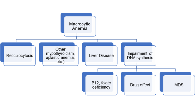
Macrocytic anemia is commonly due to folate or vitamin B12 (cobalamin) deficiency.1 Deficiency in these vitamins can be related broadly to poor intake, poor absorption, or drug interference. In patients with chronic excess alcohol consumption, both intake and/or absorption of these vitamins may be affected.

Although folate deficiency is increasingly rare in many developed countries due to mandatory folate fortification of flour and uncooked-grain, alcohol use can be associated with malnourishment severe enough to causes folate deficiency. In addition, alcohol itself can alter folate metabolism and absorption.  More specifically, chronic alcohol consumption has been shown to be associated with decreased folate absorption by the small intestine, altered intrahepatic processing and distribution between the systemic and enterohepatic folate circulations as well as increased folate urinary excretion. 2 Though uncommon,3 alcohol can also be associated with a food B12 malabsorption process, whereby despite adequate intake, B12 is not released or absorbed from food. 4

But what if serum folate and B12 levels return as normal in our patient with macrocytosis? It turns out that alcohol consumption, independent of folate or B12 deficiency, may also cause macrocytosis. 5 Though the exact mechanism is unknown, it may be related to alcohol’s direct toxicity or that of its metabolites; alcohol is oxidized to acetaldehyde, which affects membranes of red blood cells (RBCs) and their precursors by forming adducts with erythroid proteins,6 and interfering with cell division.7 Interestingly, alcohol-related macrocytosis may appear before anemia is detected and can resolve within 2-4 months of abstinence.

In addition to alcohol, cirrhosis itself may be associated with macrocytic anemia caused by lipid deposition on RBC membranes.1

See also a related pearl at  www.Pearls4Peers.com

References

  1. Hoffbrand V, Provan D. ABC of clinical haematology: macrocytic anaemias. BMJ 2011;314(7078):430–430. https://www.ncbi.nlm.nih.gov/pubmed/9040391
  2. Medici V, Halsted CH. Folate, alcohol, and liver disease. Mol Nutr Food Res 2013;57(4):596–606. https://www.ncbi.nlm.nih.gov/pubmed/23136133
  3. Bode C, Bode CJ. Effect of alcohol consumption on the gut. Best Pract Res Clin Gastroenterol [Internet] 2003;17(4):575–92. https://www.sciencedirect.com/science/article/pii/S1521691803000349
  4. Dali-Youcef N, Andrès E. An update on cobalamin deficiency in adults. QJM 2009;102(1):17–28. https://academic.oup.com/qjmed/article/102/1/17/1502492
  5. Savage DG, Ogundipe A, Allen RH, Stabler SP, Lindenbaum J. Etiology and diagnostic Evaluation of macrocytosis. Am J Med Sci [Internet] 2000;319(6):343–52. http://dx.doi.org/10.1016/S0002-9629(15)40772-4 https://www.ncbi.nlm.nih.gov/pubmed/10875288
  6. Latvala J, Parkkila S, Melkko J, Niemelä O. Acetaldehyde adducts in blood and bone marrow of patients with ethanol-induced erythrocyte abnormalities. Mol Med 2001;7(6):401–5. https://www.ncbi.nlm.nih.gov/pubmed/11474133
  7. Wickramasinghe SN, Malik F. Acetaldehyde causes a prolongation of the doubling time and an increase in the modal volume of cells in culture. Alcohol Clin Exp Res 1986;10(3):350–4. https://www.ncbi.nlm.nih.gov/pubmed/3526962


Contributed by Kim Schaefer, Harvard medical student, Boston, MA

Liked this post? Sign up under MENU and catch future pearls right into your inbox!


Why does my patient with alcoholic cirrhosis have macrocytic anemia?

My patient with anemia has an abnormally high mean red blood cell corpuscular volume (MCV). What conditions should I routinely consider as a cause of his macrocytic anemia?

Anemia with mean corpuscular volume (MCV) above the upper limit of normal (usually ≥ 100 fL) is considered macrocytic anemia. The numerous causes of macrocytic anemia can be divided into major categories (1,2) (Figure 1).

First, a reticulocyte production index should be calculated and if elevated the MCV can be above the normal range due to the large size of reticulocytes. Once high MCV is not thought to be related to reticulocytosis, the majority of macrocytic anemias can be categorized according to one of two major mechanisms: 1. Liver disease; and  2. Impairment of DNA synthesis, which includes nutritional deficiencies (folate, B12), drug effect (e.g co-trimoxazole, anti-neoplastic agents and certain anti-retroviral drugs) and “idiopathic” causes (myelodysplastic syndromes).

Mild macrocytosis can also be seen in hypothyroidism and hypoproliferative anemias such as aplastic anemia.  Macrocytosis without anemia or liver disease can also be a manifestation of heavy alcohol intake.

Macrocytic anemia in liver disease is due to excess lipid deposition in the red blood cell (RBC) membrane, not impairment of DNA synthesis. Enlarged RBCs are usually round and  often have a targeted appearance in liver disease; acanthocytes (spur cells) may also be present (Fig 2). In contrast, in disorders of impaired DNA synthesis, enlarged RBCs are often oval-shaped (macro-ovalocytes) (Fig 3).

Other common abnormalities seen with macrocytic anemia include hypersegmented neutrophils (eg, induced by B12 or folate deficiency), and in the case of myelodysplastic syndromes, hypogranulated neutrophils and Pelger-Huet neutrophil abnormalities.

Bonus pearl: Did you know that the MCV unit, fL, stands for femtoliters or 1/1,000,000,000,000,000 L? 

macroalgo

Figure 1. Major causes of macrocytic anemia. MDS: myelodysplastic syndrome.


Macrocytic_Anemia_Figure 1

Fig 2. Round macrocytes with targeting and abundant acanthocytes (spur cells) in a patient with hepatic cirrhosis.


Macrocytic_Anemia_Figure 2

Fig 3. Oval macrocytes in a patient with large granular cell leukemia and an MCV of 125 fL who received cyclophosphamide.

References

  1. Ward PC. Investigation of Macrocytic Anemia. Postgrad Med 1979; 65: 203-207. https://www.ncbi.nlm.nih.gov/pubmed/368738
  2. Green R, Dwyre DM. Evaluation of macrocytic anemias. Semin Hematol 2015; 52: 279-286. https://www.sciencedirect.com/science/article/abs/pii/S0037196315000554


Contributed by Tom Spitzer, MD, Director of Cellular Therapy and Transplantation Laboratory, Massachusetts General Hospital, Boston, MA.

My patient with anemia has an abnormally high mean red blood cell corpuscular volume (MCV). What conditions should I routinely consider as a cause of his macrocytic anemia?

Which non-pharmacological approaches may help symptoms of orthostatic hypotension in my patient with autonomic insufficiency?

A number of simple measures to help reduce the symptoms of neurogenic orthostatic hypotension (nOH) in susceptible patients have been recommended.1

  • Blood volume repletion (a minimum of 64 oz or 2L of water intake daily), depending on cardiac status. In addition, rapid consumption (within 5 min) of 16 oz or 500 ml of water can raise blood pressure by 30 mmHg for about an hour. It’s worth noting that liquids other than water (eg, water plus salt) do not provide the same BP response, likely due to water-induced hypo-osmolar reflex in the portal circulation.2,3
  • Increase salt intake if possible (eg, add 1-2 teaspoons or 2.3-4.6 g of salt per day), as many patients with nOH have an inadequate salt intake.
  • Improve physical conditioning that is not gravitationally challenging (eg, stationary recumbent bicyle, rowing machine).
  • Avoid increased core body temperature (eg hot tubs, prolonged hot showers).
  • Head-up position while sleeping through use of a wedge under the mattress or blocks under the head of the bed so that the head is 6-9 inches (15-23 cm) higher than the feet. This is to minimize nocturnal supine hypertension which can cause pressure diuresis and volume depletion.
  • Compressive garments, preferably either an abdominal binder or thigh high stockings when erect; knee high stocking are not likely to be effective.
  • Smaller, more frequent,  meals not high in carbohydrates in patients with postprandial hypotension.
  • Dietary supplementation with B12 or iron, if deficient.


Liked this post? Download the app on your smart phone and sign up below to catch future pearls right into your inbox, all for free!

Subscribe to Blog via Email

Enter your email address to subscribe to this blog and receive notifications of new posts by email.


References

  1. Gibbons CH, Schmidt P, Biaggioni I, et al. The recommendations of a consensus panel for the screening, diagnosis, and treatment of neurogenic orthostatic hypotension and associated supine hypertension. J Neurol 2017;264:1567-1582. https://www.ncbi.nlm.nih.gov/pubmed/28050656
  2. Jordan J, Shannon JR, Black BK, et al. The pressor response to water drinking in humans: a sympathetic reflex? Circulation 101:504-9. http://circ.ahajournals.org/content/101/5/504.long
  3. Raj SR, Biaggioni I, Black BK, et al. Sodium paradoxically reduces the gastropressor response in patients with orthostatic hypotension. Hypertension 2007;48:329-334. https://www.ncbi.nlm.nih.gov/pubmed/16785332

Disclosures: The listed questions and answers are solely the responsibility of the author and do not necessarily represent the official views of Massachusetts General Hospital, Harvard Catalyst, Harvard University, its affiliate academic healthcare centers, or its contributors. Although every effort has been made to provide accurate information, the author is far from being perfect. The reader is urged to verify the content of the material with other sources as deemed appropriate and exercise clinical judgment in the interpretation and application of the information provided herein. No responsibility for an adverse outcome or guarantees for a favorable clinical result is assumed by the author. Thank you!

Which non-pharmacological approaches may help symptoms of orthostatic hypotension in my patient with autonomic insufficiency?

Can oral candidiasis be symptomatic without actual pseudomembranes or “thrush”?

Yes!  Although we often associate oral candidiasis with thrush or pseudomembranous white plaques, another common form of oral candidiasis seen in hospitalized patients is “acute atrophic candidiasis” (AAC), also referred to as “antibiotic sore mouth” because of its association with use of broad spectrum antibiotics (1,2). 

Despite the absence of thrush, patients with AAC often have erythematous patches on the palate, buccal mucosa and dorsum of the tongue. Common symptoms include burning sensation in the mouth (especially with carbonated drinks in my experience), dry mouth and taste buds “being off” (2).  

Aside from antibiotics, other predisposing factors for AAC include corticosteroids, HIV disease, uncontrolled diabetes mellitus, iron deficiency anemia, and vitamin B12 deficiency.

So next time you see a hospitalized patient with new onset sore, burning mouth that wasn’t present on admission, think of antibiotic sore mouth!

Liked this post? Sign up under MENU and catch future pearls right into your inbox!

References

1. Stoopler ET, Sollecito TP. Oral mucosal diseases. Med Clin N Am 2014;98:1323-1352. https://www.ncbi.nlm.nih.gov/pubmed/25443679

2. Millsop JW, Fazel N. Oral candidiasis. Clin Derm 2016;34:487-94. https://www.ncbi.nlm.nih.gov/pubmed/27343964

Can oral candidiasis be symptomatic without actual pseudomembranes or “thrush”?